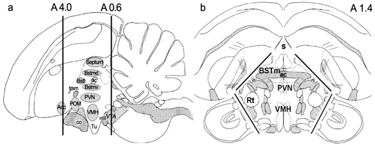
Figure 6.
The schematic drawings of dissected hypothalamic–septal region: (a) sagittal, (b) coronal section. The anterior and posterior coordinates of the dissection of a coronal slide were A4.0 and A0.6. The black lines on (b) represent the included hypothalamic and septal regions with the removal of tectum and pallial structures.

