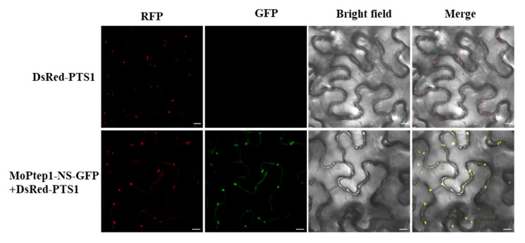
Figure 6.
Subcellular localization of MoPtep1 in the peroxisomes in N. benthamiana leaf cells. Confocal images were taken from N. benthamiana leaf cells infiltrated by EHA105 containing the corresponding constructs at 36–48 h post inoculation (hpi). DsRed-PTS1 fusion construct was the peroxisome marker. Scale bars = 10 μm.

