Figure 1.
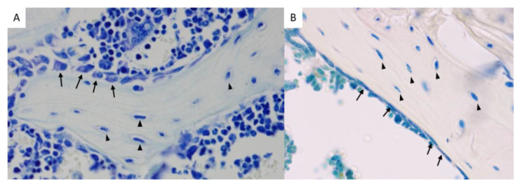
(A) Toluidine blue staining of a mouse femur showing osteoblasts (arrows) and osteocytes (triangles) (female, 8 weeks of age, original magnification 400×). (B) TRAP and toluidine blue staining of a mouse tibia, showing bone lining cells (arrows) and osteocytes (triangles) (female, 18 weeks of age, original magnification 400×).

