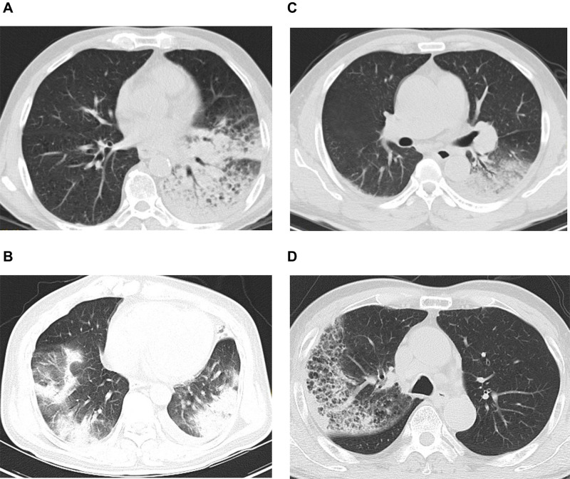
Figure 1.
(A) Chest computed tomography (CT) of Case 1: consolidation in left lower lung and a small amount of pleural effusion on the left. (B) Chest CT of Case 2: large exudative consolidation foci in both lungs, mainly in the lower lobe, and a small amount of pleural effusion on both sides. (C) Chest CT of Case 3: consolidation in both lower lungs, mainly on the left and bilateral pleural effusion. (D) Chest CT of Case 4: right lung consolidation and ground-glass shadow.

