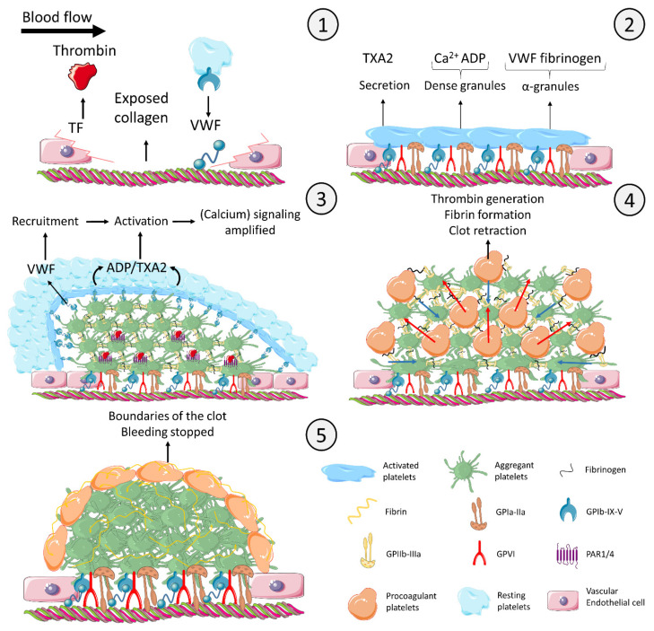
Figure 1.
Model of time-and agonist-dependent formation of the thrombus and diversification of platelets at the site of injury. Panel 1. Initiation: The injured endothelial cells express TF and secrete VWF. The subendothelial collagen becomes exposed. Initial thrombin is produced due to TF expression. Platelet GPIb-IX-V complex interacts with the VWF deposited on collagen. Panel 2. Secretion and shape change: Platelets bind to collagen via the receptor GPIa-IIa. Even though GPIb-IX-V and GPIa-IIa trigger intracellular signaling, platelet activation is mainly mediated by the collagen receptor GPVI. Activated platelets synthesize and secrete TXA2. Dense granules containing Ca2+ and ADP and α-granules containing VWF and fibrinogen are secreted. Panel 3. Aggregation and endoluminal thrombus growth: Platelets aggregate by means of fibrinogen bridging activated GPIIb-IIIa receptors (integrin αIIbβ3). Secreted TXA2 and ADP activate platelets that are recruited into the growing thrombus by VWF secreted from α-granules. Initial thrombin generation in combination with collagen further activates platelets in the thrombus core. Panel 4. Procoagulant platelets: As the thrombus is growing and the platelets become more and more activated, the increasing cytosolic level of calcium eventually creates procoagulant platelets. Procoagulant platelets downregulate their GPIIb-IIIa receptor, are coated with fibrinogen and other adhesive proteins to be retained in the thrombus, and express negatively charged phospholipids binding coagulation complexes, which localize and enhance thrombin generation. Thrombin converts fibrinogen into fibrin, which consolidates the platelet clot. In parallel, the outside-in signaling of aggregating platelets inducing clot retraction (blue arrows) squeezes procoagulant platelets out of core of the thrombus (red arrows). Panel 5. Termination: The final configuration completely stops the bleeding and defines the boundaries of the clot to avoid any undesired expansion. The figure uses modified images from Servier Medical Art under a Creative Commons Attribution 3.0 Unported License (http://smart.servier.com, accessed on 31 January 2021). This figure is inspired from [14,15,16,17,18]. Legend: ADP: adenosine diphosphate; Ca2+: calcium; GP: glycoprotein; PAR: protease activated receptor; TF: tissue factor; TXA2: thromboxane A2; VWF: von Willebrand factor.

