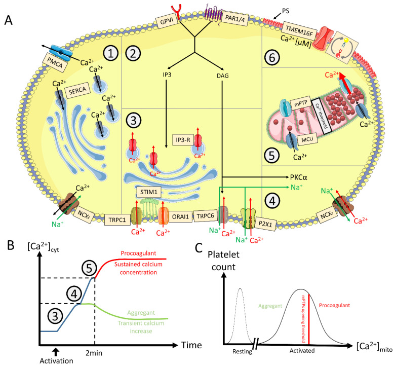
Figure 2.
Calcium mobilization mechanisms and intracellular profile leading to procoagulant COAT platelets. Panel (A). Cytosolic calcium regulation. 1. Downregulation: The intracellular level of calcium (Ca2+) is negatively regulated (Ca2+ in black) by PMCA and the forward mode of NCX (Ca2+ efflux towards the extracellular space) respectively compartmentalized by SERCA in intracellular stores (granules, DTS) and MCU in mitochondria. 2. Initial activation: Engagement of GPVI (by collagen) and PAR1/4 (by thrombin) mediate IP3 and DAG production. 3. Internal Ca2+ storage: IP3 triggers the release of Ca2+ from the internal stores through its receptor while the stores are refilled by ORAI1 mediated by STIM1 (Ca2+ sensor). TRPC1 is either agonist or storage depletion dependent and increases cytosolic Ca2+ and sodium (Na+) when activated. TRPC6 activation is mediated by DAG. ATP released from granules activates the P2X1 receptor increasing cytosolic Ca2+ and Na+. 4. NCX reversing: Increased cytosolic Na+ and PKCα contribute to the reverse mode of NCX. 5. Mitochondrial permeation: Cytosolic Ca2+ is transferred into mitochondria through MCU. At a given threshold, and in some platelets, mitochondrial Ca2+ triggers mPTP opening, releasing an important amount of Ca2+ in the cytosol. 6. PS exposure: Sustained micromolar levels of cytosolic Ca2+ are necessary to activate low Ca2+-sensitive actors such as calpain and TMEM16F. The latter scrambles membrane phospholipids inducing the expression of PS in the outer part of the cytosplamic membrane. Panel (B). Model of stepwise Ca2+ mobilization. After initial [Ca2+]cyt increase, some platelets do not trigger NCX reversing, and will remain aggregant decreasing their [Ca2+]cyt (green). Other platelets undergo NCX reversing, thus further increasing [Ca2+]cyt. Platelets become procoagulant (red) approx. 2 min after activation, when Ca2+ concentrations in cytoplasm and mitochondria reach a threshold level to induce mitochondrial depolarization, supramaximal [Ca2+]cyt, and subsequent PS exposure. Panel (C). Mitochondrial Ca2+ profile at the population level. The level of accumulated mitochondrial Ca2+ depends on previous activation of the Ca2+ mobilization sources following platelet activation. Mitochondria of a platelet subpopulation accumulate enough Ca2+ to open their PTP and induce a procoagulant response in those platelets. The figure uses modified images from Servier Medical Art under a Creative Commons Attribution 3.0 Unported License (http://smart.servier.com, accessed on 31 January 2021). This figure is inspired from [22,26,27]. Legend: [Ca2+]cyt/mito: cytosolic/mitochondrial calcium concentration; DAG: diacylglycerol; DTS: dense tubular system; IP3: inositol trisphosphate; GP: glycoprotein; PAR: protease activated receptors; MCU: mitochondrial calcium uniporter; NCXf/r: sodium calcium exchanger forward or reverse mode, respectively; PMCA: plasma membrane calcium ATPase; PS: phosphatidylserine; mPTP: mitochondrial permeability transition pore; SERCA: sarco-endoplasmic reticulum calcium ATPase; STIM: stromal interaction molecule; TMEM: transmembrane; TRPC: transient receptor potential C.

