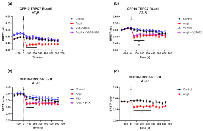
Figure 5.
Modulation of BRET ratio in response to inhibition of specific G-protein signaling and loss of β-arrestins. (a–c) HEK293 cells were co-transfected with plasmids encoding AT1R (500 ng) and GFP10-TRPC7-RLucII (75 ng). Cells were pre-incubated 10 min with YM-254890 (Gαq inhibitor; 1 µM) (a), Y27632 (ROCK inhibitor; 10 µM) (b) or 24 h with Pertussis Toxin (PTX, Gαi/o inhibitor; 100 ng/mL) (c) before BRET measurement and stimulation with AngII (1 µM) or vehicle as a control. BRET signal was measured for 10 min. (d) βArr KO HEK293 cells were co-transfected with plasmids encoding AT1R (500 ng) and GFP10-TRPC7-RLucII (75 ng) and were respectively stimulated with AngII (1 µM) or vehicle as control. BRET signal was measured for 10 min. Each data set represents the mean of three independent experiments, which were each performed in triplicate, and expressed as the mean ± S.E.M. Statistical analyses were performed using a two-way ANOVA with multiple comparisons, followed by a Sidak’s post-hoc test. * p < 0.05, ** p < 0.01 for control vs. AngII; $$ p < 0.01 for Y27632 vs. AngII + Y27632; and † p < 0.05 for PTX vs. AngII + PTX.

