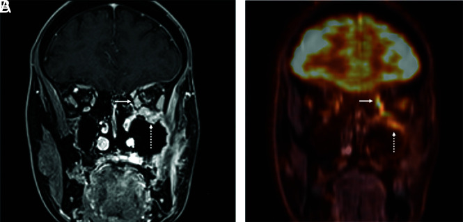
FIG 3.
PET/MR imaging to evaluate perineural spread of adenoid cystic carcinoma. Coronal T1WI postgadolinium (A) and coronal fused PET/MR imaging (B) of a patient with a history of left maxillary sinus adenoid cystic carcinoma status post resection and radiation. On this initial posttreatment PET/MR imaging, the enhancing tissue along the thickened extraocular muscles (arrow) and infraorbital foramen (dashed arrow) of the left orbit corresponds to areas of increased FDG uptake and increased metabolic activity. The site was scored as NI-RADS 3 at consensus. The patient underwent orbital exenteration, with surgical pathology positive for perineural spread of tumor.

