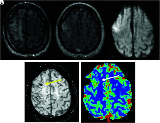
FIG 2.
A 62-year-old man with a history of lymphoplasmacytic lymphoma treated with resection and 40 Gy of whole-brain radiation therapy presented with seizure, left hemiparesis, and speech impairment. A and B, A FLAIR hyperintense region associated with gyriform enhancement and subcortical WM edematous change in the right frontal and parietal lobes. C and D, DWI and SWI show restricted diffusion (ADC not shown) and SWI hypointensity in the same area (yellow arrow). E, DSC MR imaging shows an increase in rCBV in the corresponding area (white arrow). The patient recovered incompletely, with remaining left hemiparesis.

