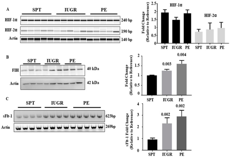
Figure 2.
HIF gene regulation, expression of Factor inhibiting HIF (FIH) and Expression of the HIF regulated gene sFlt-1 in placental tissues. (A) mRNA of normal (n = 13), IUGR (n = 16), and PE (n = 19) pregnancies was probed for HIF-1α and 2α transcript levels by RT-PCR. (B) Placental lysates from normal (n = 13), IUGR (n = 16), and PE (n = 19) pregnancies were probed for FIH expression. Immunoblotting for β-actin was used to control for loading. (C) mRNA of normal (n = 13), IUGR (n = 16), and PE (n = 19) pregnancies was probed for sFlt-1 transcript levels by RT-PCR. β-actin transcript mRNA levels were assessed as a control (A and C). The densitometric value of one normal pregnancy control was normalised to one and changes observed in the other samples shown as a fold change compared to this reference sample. Representative blots/gels and densitometric analysis of expression are shown. Data represent mean ±SEM. p values as compared to normal pregnancy are indicated. NS = Not significant. Full gels for immunoblots can be found in SF2.

