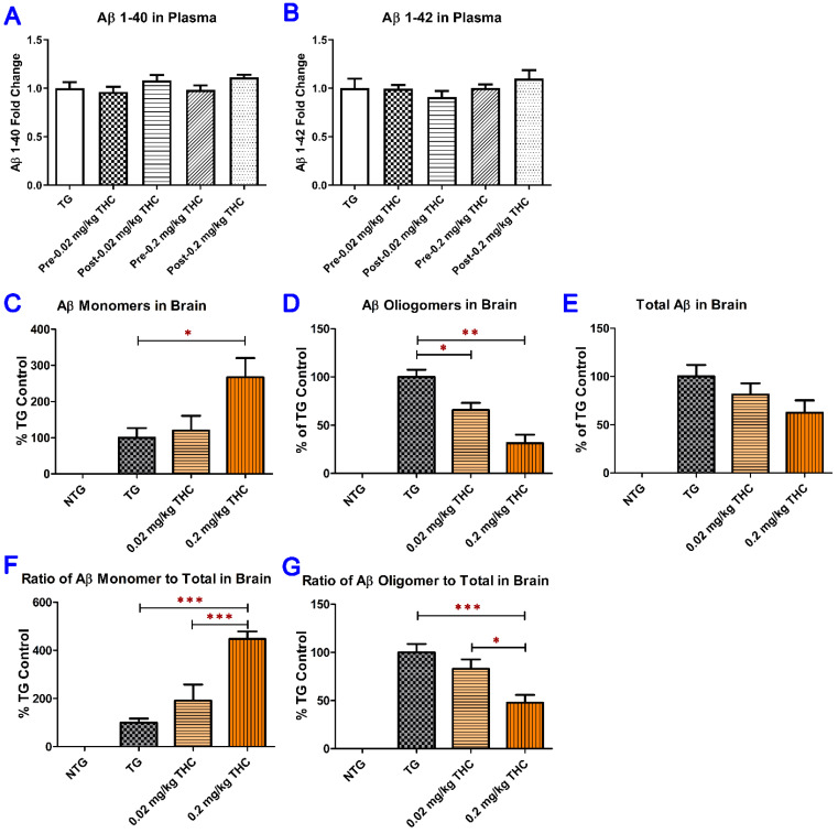
Figure 6.
Determination of both soluble and insoluble Aβ1–40 (A) and Aβ1–42 (B) levels in the plasma using ELISA. Plasma samples were collected from the APP/PS1 mice before the start and after the 3-month THC treatment. No significant difference in soluble and insoluble Aβ1–40 and Aβ1–42 levels in plasma was found among all treatment groups and between the baseline and post-treatment levels (N = 7 for the control NTG group, N = 6 for the control TG and 0.2 mg/kg THC groups, and N = 8 for the 0.02 mg/kg THC group). Determination of Aβ monomers (C), Aβ oligomers (D), and total Aβ (E) in mouse brain tissue using the semi-quantitative western blotting and the total Aβ normalized Aβ monomer (F) and oligomer levels (G). The total Aβ normalized Aβ monomer (or oligomer) level was calculated as the ratio of Aβ monomer (or oligomer) level to total Aβ level. Treatment with 0.2 mg/kg THC significantly increased the Aβ monomer level and decreased Aβ oligomer level compared with the control treatment in TG mice. Data are expressed as mean ± SD (N = 6 for each study group). SD is denoted by the error bars. * p < 0.05, ** p < 0.01, and *** p < 0.001 compared between the control NTG mice, control APP/PS1 mice, and APP/PS1 mice treated with 0.02 and 0.2 mg/kg THC using one-way ANOVA followed by the Tukey–Kramer post hoc multiple comparison test.

