Figure 8.
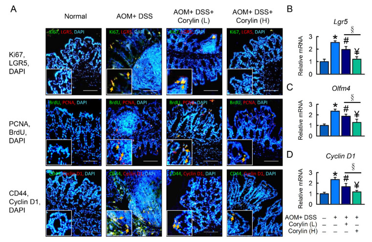
Corylin affects LGR5 and proliferation in AOM/DSS-induced-colitis-associated colorectal cancer mice. (A) Top line: Double immunofluorescence staining of Ki67 (green), LGR5 (red), and DAPI (blue) in colon. The staining for Ki67/LGR5 from colon expressing both markers is identified. Middle line: Double immunofluorescence staining of BrdU (green), PCNA (red), and DAPI (blue) in colon. The staining for BrdU/PCNA from colon expressing both markers is identified. Bottom line: Double immunofluorescence staining of endogenous CD44 (green), cyclin D1 (red), and DAPI (blue) in colon. The staining for CD44/cyclin D1 from colon expressing both markers is identified. DAPI (blue). Yellow arrows highlight positive staining. Scale bar: 100 μm. (B–D) mRNA expression of Lgr5, Olfm4, and Cyclin D1 as determined by qRT-PCR. Scale bar: 100 μm. Results represent mean ± SEM. * p < 0.05, Normal compared with AOM/DSS; # p < 0.05, AOM/DSS compared with AOM/DSS + Corylin (L); ¥ p < 0.05, AOM/DSS compared with AOM/DSS + Corylin (H); § p < 0.05, DSS + Corylin (L) compared with DSS + Corylin (H). AOM, azoxymethane; DSS, dextran sodium sulfate; LGR5, leucine-rich repeat-containing G-protein-coupled receptor 5; PCNA, proliferating cell nuclear antigen; BrdU, bromodeoxyuridine (5-bromo-2′-deoxyuridine; DAPI, 4′,6-diamidino-2-phenylindole.

