Figure 2.
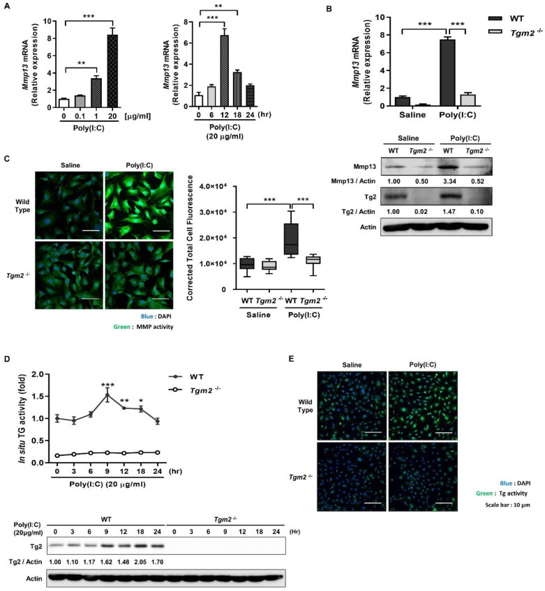
Tgm2-deficient MDFs exhibit reduced Mmp-13 expression in response to poly(I:C) treatment. (A) MDFs were treated with various concentrations of poly(I:C), and mRNA levels of Mmp13 were measured after 12 h using qRT-PCR (left, n = 3). Mmp13 mRNA levels of MDFs treated with 20 µg/mL of poly(I:C) were monitored for 24 h (right, n = 3). (B) Mmp13 mRNA level (upper panel) and Mmp13 protein (lower panel) of wild-type and Tgm2−/− MDFs were measured using qRT-PCR and western blotting, respectively, after treatment with 20 µg/mL of poly(I:C) for 12 h. (n = 3) (C) Intracellular MMPs activity of wild-type and Tgm2−/− MDFs was visualized after treatment with 20 µg/mL of poly(I:C) for 12 h using DQ™-collagen I (left) and quantitated by measuring corrected total cell fluorescence using Image J (right, n = 11). (D) Wild-type and Tgm2−/− MDFs were treated with poly(I:C) (20 µg/mL for 12 h). Intracellular TG activity (upper panel) and Tg2 protein level (lower panel) were monitored for 24 h using biotinylated pentylamine (BP) incorporation assay (n = 3) and western blotting. Intracellular TG activity was visualized by detection of BP incorporated proteins using Streptavidin-FITC (E). All data are represented as mean ± SEM. Statistical significance was tested by one-way ANOVA by Dunnett’s post-test (A) or two-way ANOVA by Turkey’s post-test (B–D). *, p < 0.05; **, p < 0.01; ***, p < 0.001.

