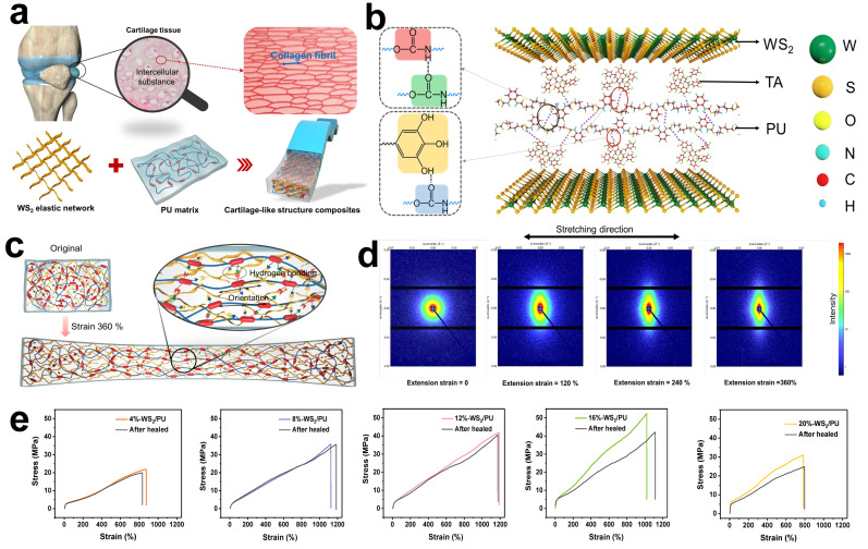
Figure 8.
(a) Schematic illustrations of a cartilage structure, intercellular substance of cartilage tissue and the nanostructure of composite consisting of hydrogen-bonded interwoven network of 2D WS2 and PU matrix. (b) Schematics of the dynamic noncovalent bonding interaction between PU and interwoven network of 2D WS2. (c) Schematic illustrations of the nanostructure of the original sample and stretching sample. (d) 2D SAXS images of the 16 wt% TA-WS2/PU with different tensile strain during uniaxial stretching process. (e) Mechanical self-healing performance of PU composites filled with different contents of TA-WS2. Reproduced with permission [177]. Copyright 2021 Springer Nature.

