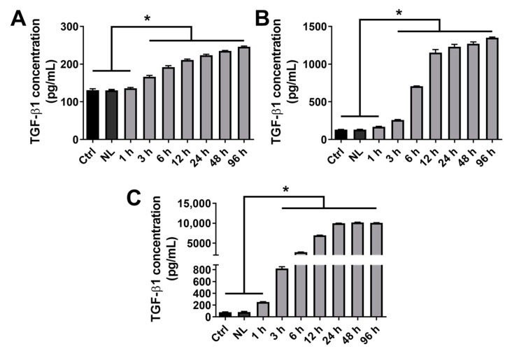
Figure 3.
Kinetics of TGF-β1 release from rapeseed nanoliposomes in cell culture medium. Cells were untreated or exposed to 100 µg/mL nanoliposomes, either encapsulating or not 0.1 (A), 1 (B) or 10 (C) ng/mL of recombinant TGF-β1 from 1 h to 96 h. The release of TGF-β1 in cell culture media was quantified by ELISA. The reported data are represented as mean ± SD of at least four individual experiments. Significance is indicated as * with p < 0.01. Ctrl—control cells meaning untreated chondrocytes; ELISA—enzyme-linked immunosorbent assay; NL—empty nanoliposomes; NL-TGF—TGF-β1-loaded nanoliposomes; NL-TGF 0.1—nanoliposomes encapsulating 0.1 ng/mL of TGF-β1; SD—standard deviation; TGF-β1—transforming growth factor-β1.

