
Figure 2.
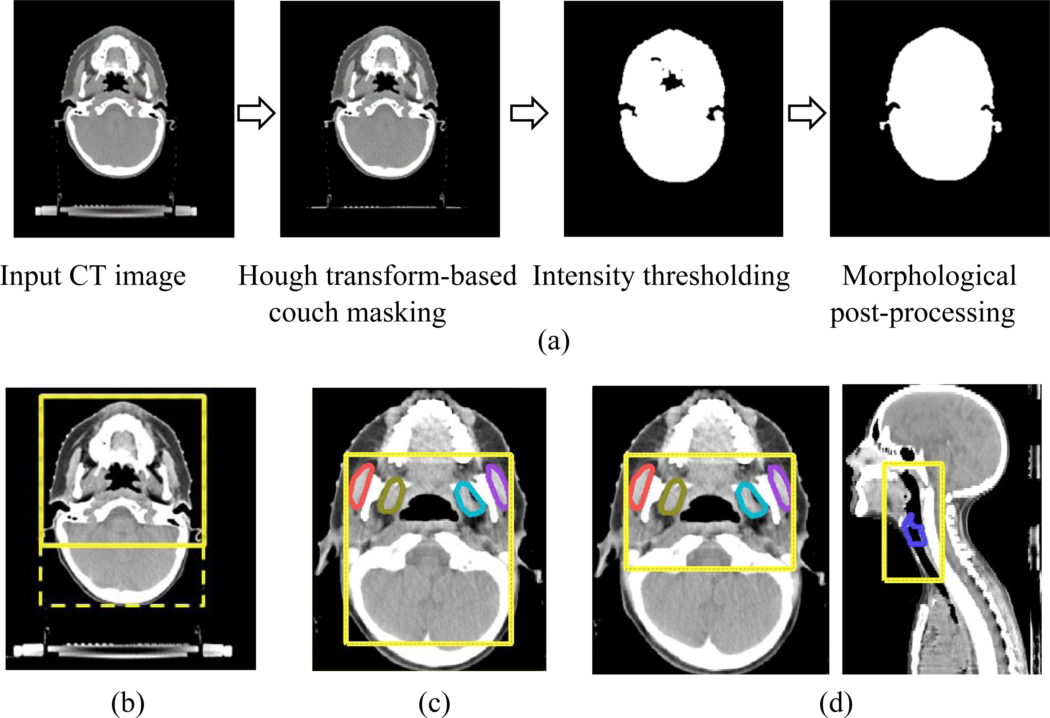
(a) Illustration of automatic method to extract patient outline in axial H&N CT scans and bounding boxes (yellow) generated sequentially to localize (b) chewing structures based on cropped patient outline (c) larynx based on previously-identified chewing structures and (d) constrictor based on previously-identified chewing structures and larynx.