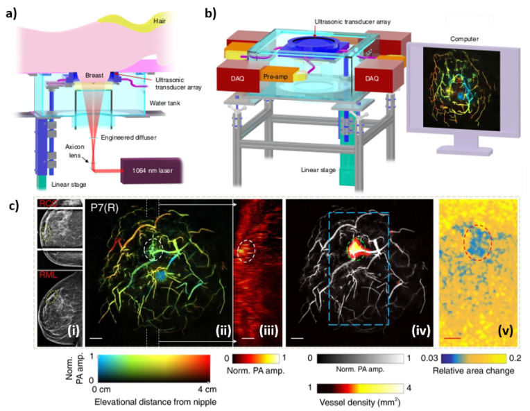
Figure 9.
(a) Overview of the SBH-PACT system. (b) Perspective view of the system with patient bed and optical components removed. DAQ: data acquisition system, Pre-amp: pre-amplifier circuits. (c) X-ray and photoacoustic (PA) images of a 44-year-old female patient with a fibroadenoma in the right breast—(i) X-ray mammograms of the affected breasts, (ii) Depth-encoded angiograms, (iii) Maximum amplitude projection images of thick slices in sagittal planes marked by white dashed lines in (ii), (iv) Automatic tumor detection on vessel density maps, and lastly (v) PA elastography images.

