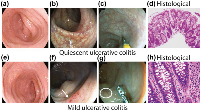
FIGURE 1.

(a–c) quiescent ulcerative colitis (UC) assessed by HD‐white light endoscopy (a); i‐scan modes 2 (b); i‐scan modes 3 (c); histology showing minimal architectural distortion of crypts and no active inflammatory infiltrate in lamina propria (d); (e, f) mild UC assessed by HD‐white light endoscopy (e); i‐scan modes 2 showing vessels with dilatation (arrow) (f); i‐scan modes 3 showing micro erosions (circle) (g); histology showing architectural distortion of crypts and focal active inflammatory infiltrate in lamina propria (h)
