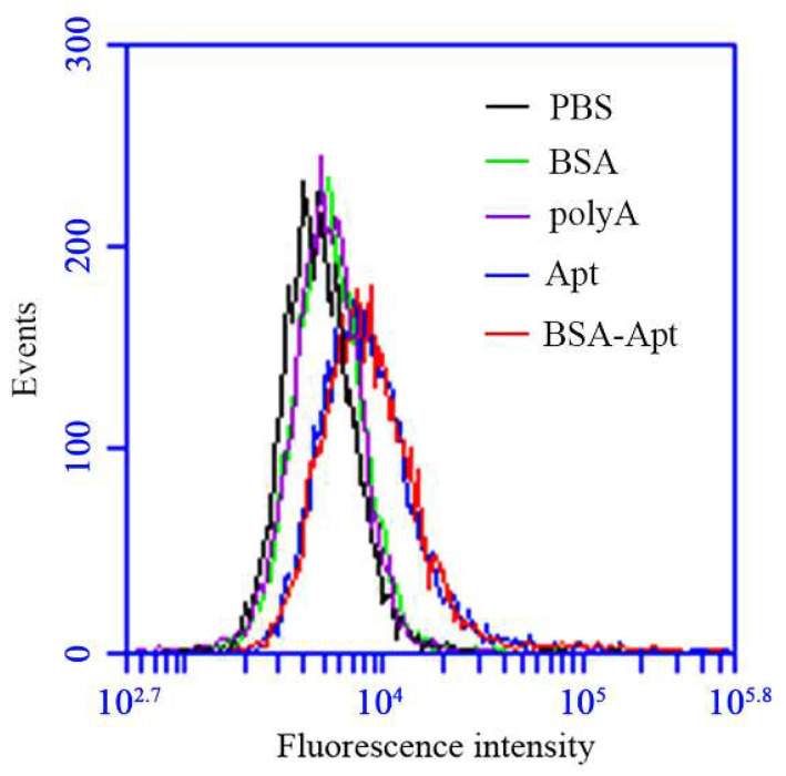
Figure 4.
The bindings of BSA, polyA, free PD-L1 aptamer, or BSA-Apt to PDL1-positive CT26 cells. CT26 cells (2 × 10⁵) were incubated with 60 pmol of FAM-labeled BSA (green), polyA (purple), free PD-L1 aptamer (blue), or BSA-Apt (red), respectively. The cells were washed with PBS and analyzed by flow cytometry.

