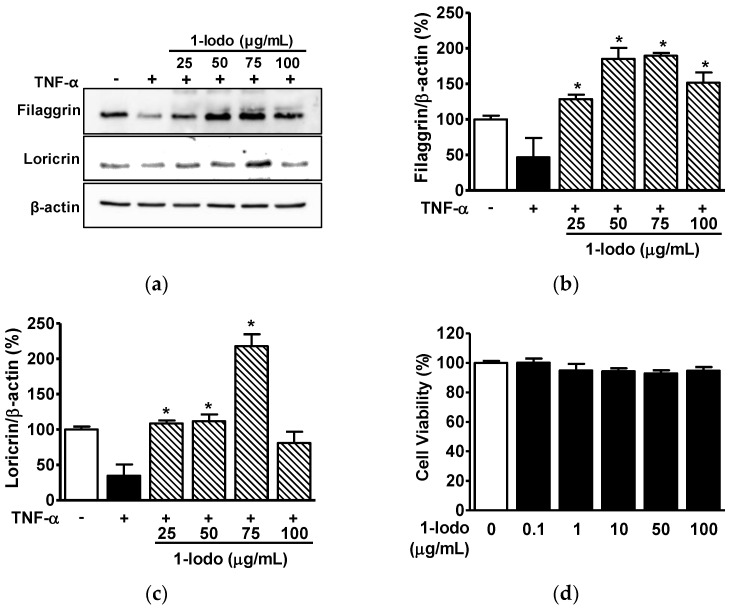
Figure 3.
Effects of 1-iodohexadecane on the expressions of skin barrier-related proteins in TNF-α-stimulated keratinocytes. (a–c) Expressions of skin barrier-related proteins. HaCaT cells were cultured at 37 °C for 36 h in DMEM in the presence or absence of TNF-α (5 ng/mL) with or without 1-iodohexadecane (1-Iodo; 25–100 μg/mL). Cell lysates were immunoblotted with the indicated antibodies. (a) Representative images. (b,c) Results for filaggrin (b; n = 3) and loricrin (c; n = 3) were obtained from panel A. (d) Cell viabilities. HaCaT cells were treated with 1-Iodo (0.1–100 µg/mL) for 36 h, and viabilities were measured using an EZ-CyTox kit (n = 5). Responses are expressed as percentages of untreated cells. The results are represented as means ± SEMs. * p < 0.05 vs. TNF-α alone-stimulated cells.

