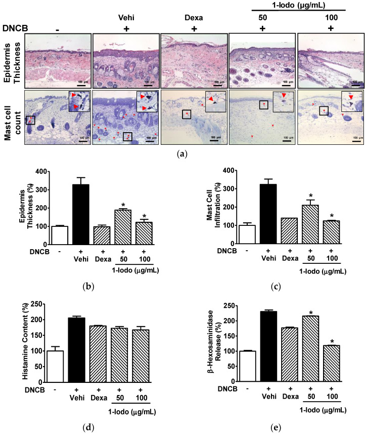
Figure 5.
Histopathological features of skin lesions and serum levels of mast cell degranulation markers in DNCB-induced mouse model treated with 1-iodohexadecane. The dorsal skins of mice were treated with 2,4-dinitrochlorobenzene (DNCB) with or without 1-iodohexadecane (1-Iodo; 50 or 100 μg/mL) for 21 days. The skin tissues were excised, fixed with 4% formaldehyde, embedded in paraffin, and sectioned as described in the Materials and Methods section. The blood was collected and centrifuged to obtain the serum as described in the Materials and Methods section. (a) Representative histological images. Sections were stained with H&E for epidermal thickness measurements or toluidine blue for mast cell counting. Red arrowheads indicate mast cells stained with toluidine. Scale bar = 100 μm. (b,c) Graphs of the results shown in panel (a). The epidermal thickness (upper images of panel (a), n = 3) and mast cell infiltration (lower images of panel (a), n = 3) were measured using Image J software. (d,e) Serum levels of histamine and β-hexosaminidase. Levels of histamine (d; n = 2) and β-hexosaminidase (e; n = 2) in the obtained sera were measured using an enzyme immunoassay. Dexamethasone (Dexa, 0.1%) in each test was used as a positive control. Values are expressed as percentages of those of non-treated controls. The results are presented as means ± SEMs. * p < 0.05 versus DNCB controls. Vehi, vehicle (100% olive oil).

