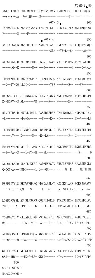
FIG. 3.
Comparison by alignment of V. parahaemolyticus (upper) and E. coli (lower) ParC amino acid sequences. Residue numbers are given for the sequence of V. parahaemolyticus. An asterisk indicates absence of a residue. Identical residues are indicated by dashes. Arrows above the amino acid sequences correspond to the orientations and positions of VGYR-1, VGYR-3, and VGYR-4 degenerated primers (Table 1) used to amplify the QRDR of V. parahaemolyticus ParC.

