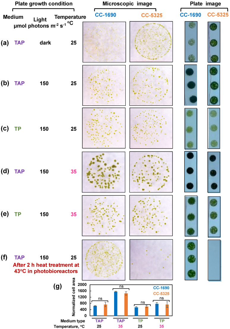
Figure 5.
CC-5325 grew better in the dark with acetate but was more heat-sensitive than CC-1690. (a–e) Algal cells grown in PBRs at 25 °C under the same condition as in Figure 2 were harvested, diluted, and spotted on agar plates, and grown under the indicated growth conditions in temperature-controlled incubators. Cultures with the same cell density were used for each spot. (f) Algal cells were heat-treated at 43 °C for 2-h in PBRs before spotting; cultures with equal volume and the same dilution were used for each spot. TAP, Tris-acetate-phosphate medium with acetate. TP, Tris-phosphate medium without acetate. The same dilution and growth duration were used for the two strains under the same condition. (a,f) Algal cultures with 1:20 dilution, about 1000 cells in 10 μL, were used for spotting. (b–e) Algal cultures with 1:100 dilution, about 200 cells in 10 μL, were used for spotting. Single algal spots were imaged using a dissect microscope after 5-day (a) or 44-h (b–f) of growth. Plate images (all 1:20 dilution) were taken after 10-day (a) or 3-days (b–f) of growth using a regular camera. Images shown are representative of three biological replicates. (g) Normalized cell area. The 44-h spotting images were analyzed by ImageJ to get cell areas which were then normalized to the number of cells spotted. Statistical analyses were performed using a two-tailed t-test assuming unequal variance by comparing with CC-1690 under the same experimental conditions. Not significant, ns. Such quantification was not performed for (a,f) because the cell areas for one of the strains were too small to quantify.

