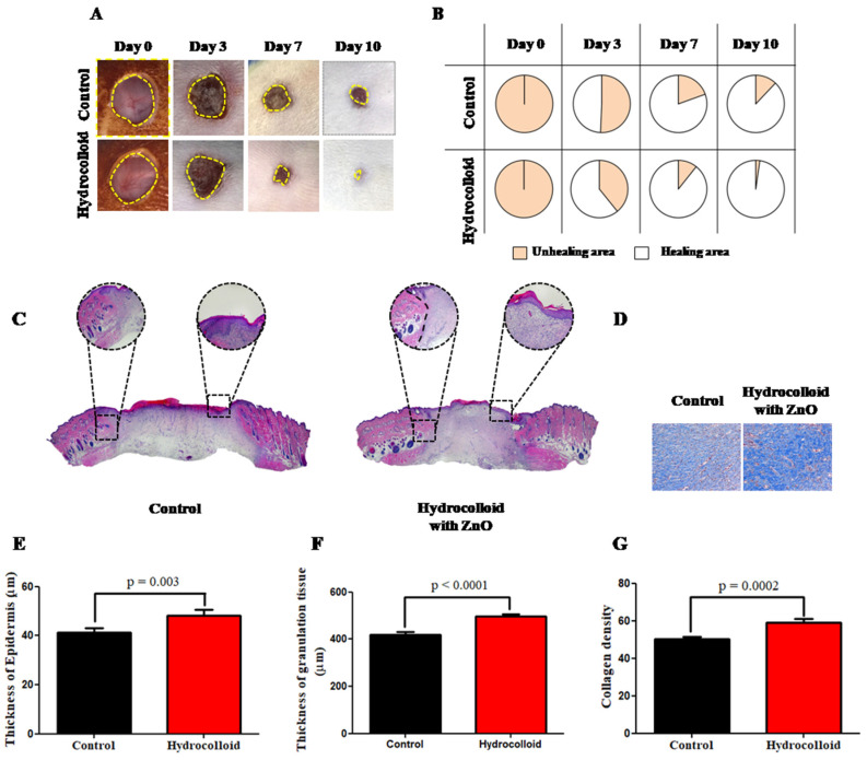
Figure 1.
A hydrocolloid patch covered with ZnO-NPs accelerates wound healing on both macroscopic and microscopic scales. (A) Representative images (1× magnification) of the wound of the hydrocolloid-with-ZnO-NPs patch and the control group throughout the healing process. (B) Wound healing closure rates of these two groups. The rates presented as a percentage of the initial wound area on day 0. (C) 4× magnification images of H&E staining on day 10 after treatment with a hydrocolloid patch covered with ZnO-NPs and without treatment. (D) 10× magnification images of MT staining on day 10. The blue color indicates the distribution of the collagen. (E) Quantified epidermis thickness gap on day 10. (F) Quantified granulation tissue gap on day 10. (G) Quantified collagen density on day 10.

