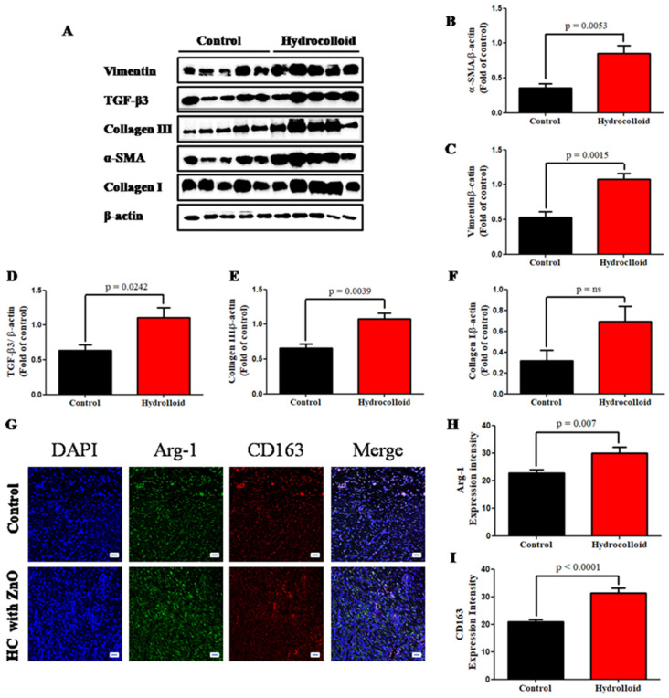
Figure 5.
A hydrocolloid patch covered with ZnO nanoparticles accelerates proliferative phases and increases the proliferative responses of wound healing. (A) Representative images of Western blotting. Quantitative densitometry analysis of proliferative response expression. (B) Alpha smooth muscle actin, α-SMA. (C) Vimentin. (D) Transforming growing factor-beta 3, TGF-β3. (E) Collagen III. (F) Collagen I. (G) Fluorescent micrographs showing cytokine staining of macrophage 2 on day 10 of the wound healing process. The quantitative density gap of macrophage 1 antigens of the two groups on day 10. (H) Arginase 1, Arg-1. (I) CD163. Scale bars are 50 µm.

