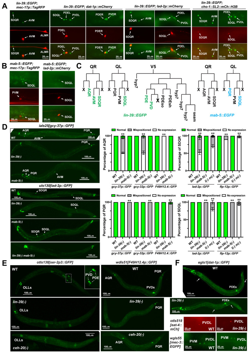
Fig 2. LIN-39 promotes neuronal fate specification in the Q and V5 lineage.

(A) The expression of lin-39 in AVM, SDQL/R, PDEL/R, and PVDL/R, indicated by the overlapping with neurotransmitter identity markers and specific fate markers (uIs115[mec-17p::TagRFP] for AVM, otIs181[dat-1p::mCh] for PDE, uIs117[lad-2p::mCh] for SDQ). (B) The expression of mab-5 in SDQL. (C) Summary of lin-39 (green) and mab-5 (cyan) expression in the descendants of Q and V5 lineages. (D) The loss of gcy-37 expression in AQR and AVM neurons in lin-39(n1760) mutants and the mispositioning of PQR in mab-5(gk670) mutants; the loss of lad-2 expression in SDQR in lin-39(n1760) mutants, the displacement of SDQL in mab-5(gk670) mutants, and the loss of lad-2 expression in both SDQs in lin-39(n1760) mab-5(e1239) mutants. The right panels show the penetrance for the loss of marker expression and cell body mispositioning. Mean ± SD for the percentage of cells showing corresponding phenotypes from three biological replicates are shown. Double asterisks indicate statistically significant difference (p < 0.01) between the mutants and the wild type in a Chi-square test. (E) The loss of ser-2 expression in PVD and PDE neurons and the loss of F49H12.4 expression in PVD in lin-39(n1760) and ceh-20(u843) mutants. (F) Dopaminergic marker dat-1 is normally expressed in PDE neurons in lin-39 mutants, but PDE shows axonal growth defects. The arrows indicate the termini of PDE axons. The expression of glutamatergic identity marker eat-4 and the PVD terminal selector gene mec-3 in PVD neurons in lin-39 mutants.
