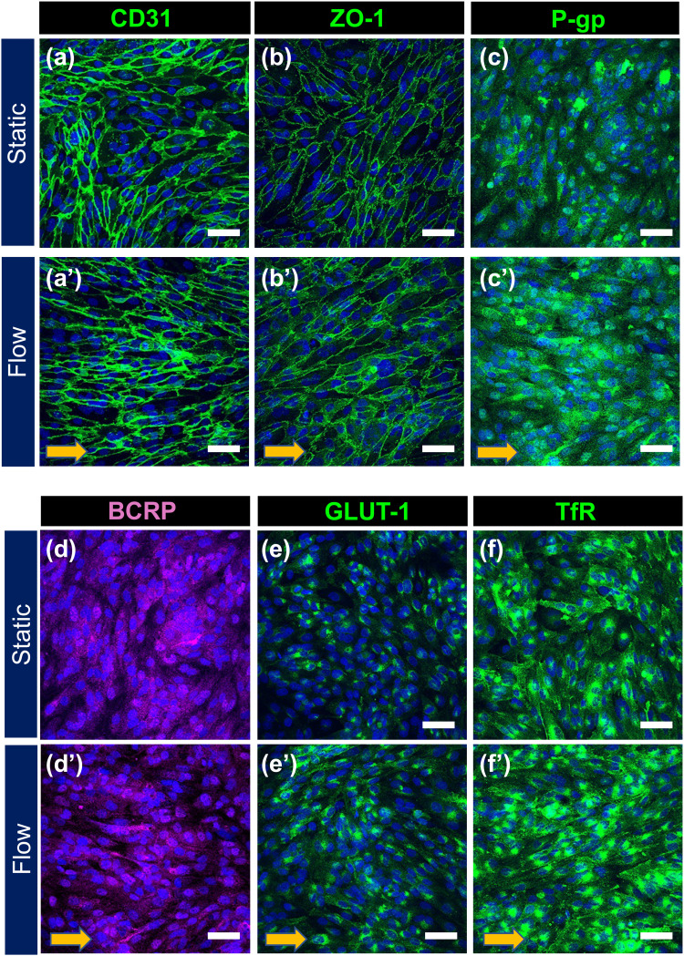
FIG. 3.
Expression of BBB marker proteins in HBMEC/ci18 cells after static and perfusion culture in the device. Confluent HBMEC/ci18 cells were cultured in the device for 72 h under static (a)–(f) or flow conditions [(a′)–(f′), 0.3 dyn/cm2]. Localization of BBB marker protein was analyzed by immunocytochemistry. Cell nuclei were counterstained with DAPI (blue). Arrows in panels (a′)–(f′) represent the flow direction. Scale bars, 50 μm.

