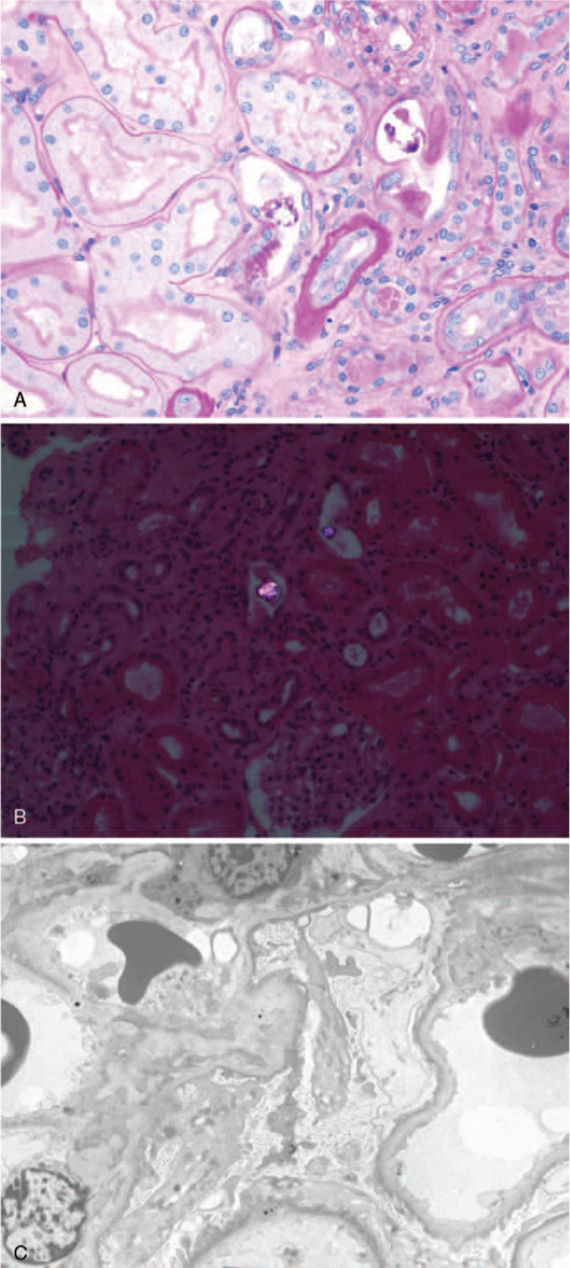
Figure 1.
Renal biopsy of a patient with oxalate nephropathy secondary to ingestion of Chaga mushroom. (A) Hematoxylin and eosin staining viewed under light microscopy showed focal acute tubular injury, deposition of calcium oxalate crystals in tubules, interstitial fibrosis, and tubular atrophy. (B) The calcium oxalate crystals demonstrated birefringence under polarized light. (C) Electron microscopy showed that there was an effacement of foot processes in podocytes.

