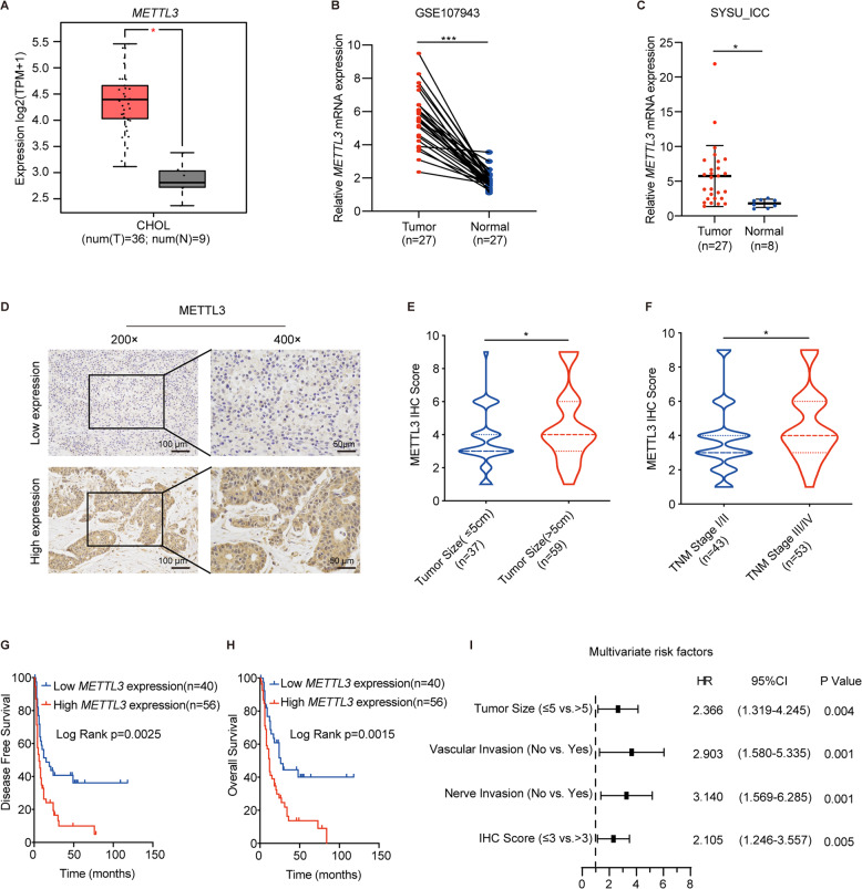
Fig. 1. Elevated METTL3 expression is correlated with poorer prognosis of ICC patients.
A METTL3 expression of ICC tumor (n = 36) and normal tissue (n = 9) in GEPIA2 database. B METTL3 expression of ICC tumor (n = 27) and normal tissue (n = 27) in public dataset GSE107943. C METTL3 expression in ICC tumor (n = 27) and adjacent normal bile duct tissue (n = 8) were measured by real-time quantitative reverse transcription polymerase chain reaction (qRT-PCR). D Representative immunohistochemistry (IHC) staining images of ICC tumors expressing low or high levels of METTL3. E Correlation analysis of METTL3 expression with tumor size (≤5 cm vs. >5 cm). F Correlation analysis of METTL3 expression with tumor, node, metastasis stages (stage I/II vs. III/IV). G Kaplan–Meier survival curves of disease-free survival (DFS) in 96 ICC patients, stratified by METTL3 IHC score (METTL3 low expression, n = 40 vs. METTL3 high expression, n = 56). The P value was calculated using the log-rank test. H Kaplan–Meier survival curves of overall survival (OS) in 96 ICC patients, stratified by METTL3 IHC score (METTL3 low expression, n = 40 vs. METTL3 high expression, n = 56). The P value was calculated using the log-rank test. I Multivariable analyses for overall survival were performed in the ICC cohort. *P < 0.05, ***P < 0.001, according to Student’s t test.

