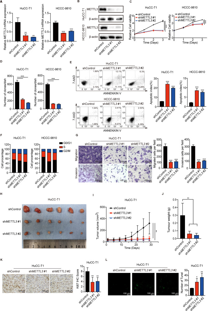
Fig. 3. METTL3 knockdown inhibits ICC progression.
A The mRNA level of METTL3 after METTL3 silencing in HuCC-T1 and HCCC-9810 cells was confirmed by RT-qPCR. B The protein level of METTL3 after METTL3 silencing in HuCC-T1 and HCCC-9810 cells was confirmed by Western blotting. C Cell growth curve of HuCC-T1 and HCCC-9810 cells transfected with METTL3 shRNA or Control. D Colony-forming assays after METTL3 silencing in HuCC-T1 and HCCC-9810 cells. E Apoptosis analysis of HuCC-T1 and HCCC-9810 cells transfected with shControl or shMETTL3. F Cell cycle analysis of HuCC-T1 and HCCC-9810 cells transfected with shControl or shMETTL3. G Cell migration ability and cell invasion ability after shMETTL3-transfection in HuCC-T1 cell. H Xenograft tumors in each group were shown. The mice were sacrificed 28 days post-injection. I Tumor growth curves after the injection of shMETTL3 and Control HuCC-T1 cells. Tumor volume was calculated every 4 days. J Tumor weight of shMETTL3 and Control groups was measured. K Representative IHC staining of Ki67 in tumors with different treatments. L Representative images of TUNEL analysis in tumors with different treatments. The results are presented as mean ± SD of three independent experiments. *P < 0.05, **P < 0.01, ***P < 0.001, according to a Student’s t test.

