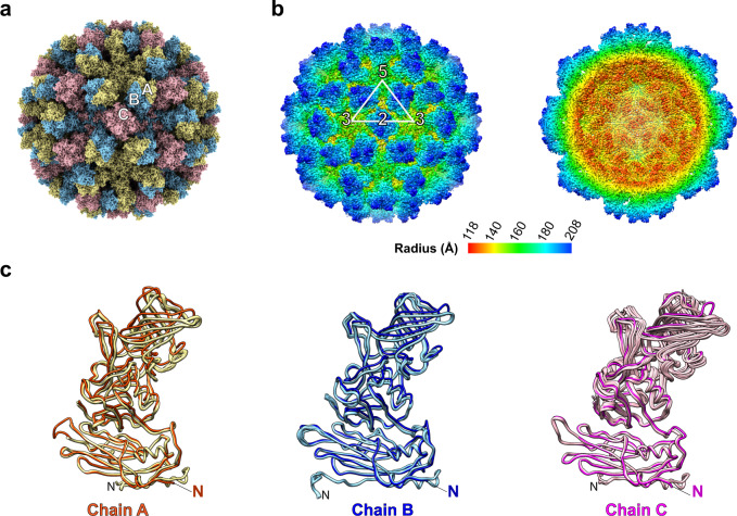
Fig. 3. Cryo-EM structure of GII.4 HOV VLP.
a GII.4 HOV VLP cryo-EM structure aligned to the crystal structure shown in Fig. 1 and viewed along the icosahedral twofold axis, shows the T = 3 symmetry of GII.4 HOV VLP in solution. The subunits A, B, and C are colored yellow, blue, and pink. b The surface and cut-away views of a GII.4 cryo-EM map colored by radial distance, showing similar particle radius as observed in the crystal structure. c Superimposition of subunit A (orange) with B (blue) or C (magenta) subunits of the cryo-EM structure with that of the VLP crystal structure (subunit A, yellow; subunit B, light blue; subunit C, pink) using the S domain. The N-terminus of VP1 in the cryo-EM and crystal structure is labeled with colored and black “N“s.

