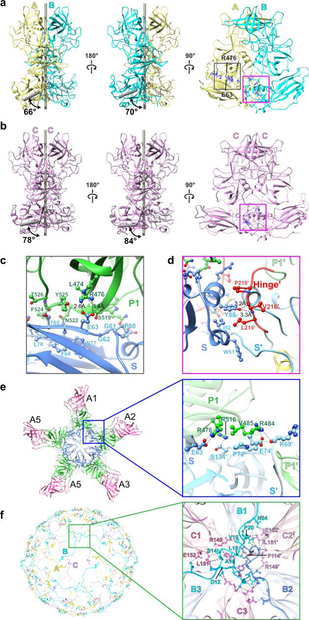
Fig. 4. Molecular interactions within and between VP1 subunits.
a, b Representative A/B and C/C dimer of the crystal structure. The angles formed by the P dimer and S domain axes are measured using Chimera, showing the bent A/B dimer with smaller angles and the flat C/C dimers with larger angles. The molecular interactions with and between VP1 are determined using Chimera and shown as blue lines. The S-P1 and S-Hinge interactions are indicated by gray and magenta boxes, respectively. c Close-up view showing the interactions between the P1 subdomain (green) and the S domain (blue). The interacting residues are shown as ball-and-stick models. d Close-up view of the interactions between the S domain (blue) and the hinge region of the neighboring subunit (red). The residues from the adjacent subunit are indicated with prime (′). e Interactions between the A1–A5 subunits at a five-fold axis. The inset shows the P1 subdomain of A1 interacting with the S domain of the A2 subunit. f The NTA network. Only residues of NTA are shown on the left panel for clarity. The inset presents how NTAs interact with each other and the S domains at a three-fold axis. The subunits B and C are colored in blue and pink, respectively.

