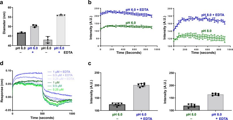
Fig. 6. Dynamic light scattering, Bis‐ANS, and BLI binding assays.
a DLS analysis shows the hydrodynamic radius increase upon removal of metal ions by chelation with EDTA at pH 6.0 and pH 8.0, indicating the rising of P domain above shell in the absence of ion at the dimeric interface. b Changes in bis-ANS binding in the absence or presence of EDTA show increased fluorescence intensity when VLPs are incubated with 20 mM EDTA, suggesting more hydrophobic surfaces of VLP bound with bis-ANS. c Stabilized fluorescence intensities measured during the last minute for each sample were averaged and presented as a bar graph. d BLI analysis of NORO-320 Fab and GII.4 HOV VLP shows that more VLPs bind to the immobilized Fab of GII.4 mAb NORO-320 in the presence of 20 mM EDTA, suggesting the exposure of mAb-binding epitope with EDTA-treatment. Data presented in each panel are means ± SE (n = 3 independent study repliates) are shown. Source data are provided.

