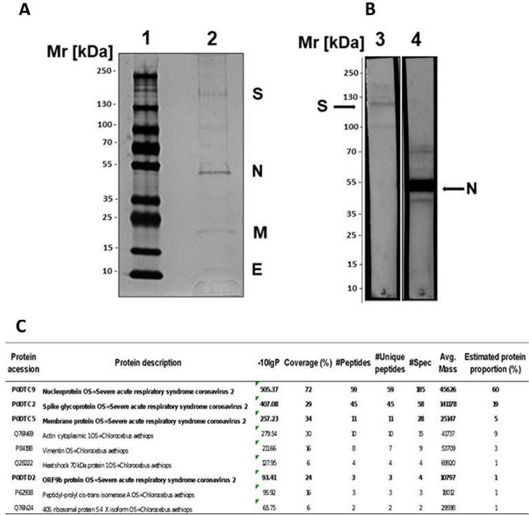
Figure 1.
Analysis of the purified and inactivated SARS-CoV-2. (A) SDS–PAGE: 4–15% gradient SDS–PAGE gel under nonreducing conditions and silver staining. (1) LMW marker—10 to 250 kDa; (2) Purified and inactivated SARS-CoV-2. (B) Western blot: samples of the virus, which were separated on 4–15% gradient SDS-PAGE gels, were blotted onto nitrocellulose membranes and incubated with monoclonal antibodies against the SARS-CoV-2 S protein (Lane 3) or anti-SARS-CoV-2N protein (Lane 4). The membranes were incubated with specific peroxidase-conjugated anti-rabbit IgG (1:5000), and the reactions were revealed using SuperSignal West Pico chemiluminescent substrate. (C) Protein identification through tandem mass spectrometry analysis (LC–MS/MS) of the inactivated SARS-CoV-2 purified antigen. Only proteins with at least 1 unique peptide, a score − log10P > 20 and a false discovery rate < 1% were accepted for identification and are depicted in the Table. The proportion of proteins was estimated based on the mass spectra counts of each identified protein. (A) and (B) were cropped, the original results are presented in Fig. S7. (B) is composed of two panels, representing two membrane strips processed separately, and a third strip containing a molecular weight marker was used do estimate the molecular sizes of bands (Fig. S7).

