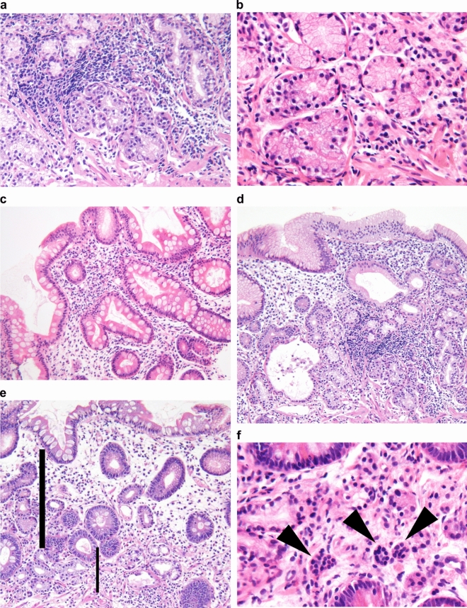
Figure 3.
Targeted histological findings of autoimmune gastritis. Oxyntic mucosal atrophy with lymphocytic infiltrates (a). Epithelium of pseudo-pyloric (b) and intestinal metaplasia (c). Diffuse lymphocyte cell infiltration, which is heavier in the deep portion than in the lamina propria (d). Increased proportion of gastric pits (bold line) induced by severe atrophy, which was defined as the proportion of gastric pits (bold line)/gastric duct (thin line) (e). Enterochromaffin-like cell hyperplasia (arrowhead) (f).

