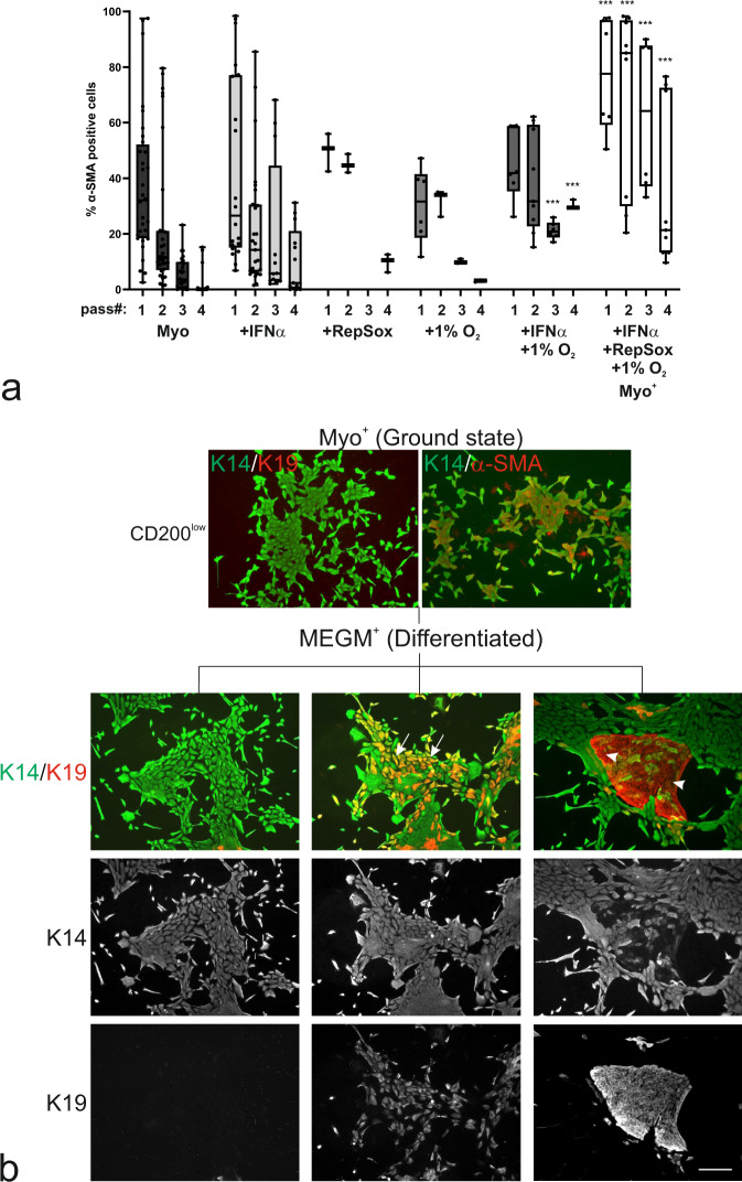
Fig. 3. Myo+ conditions support ground state of MEP progenitors poised for luminal differentiation.
a Whisker boxplot of quantification of the frequency of α-SMA-positive cells (%) in passage 1 to 4 of Trop2+/CD271high MEP cells on iHBFCCD105 feeders. In Myo medium (black), Myo medium with IFNα (light grey) or RepSox (medium grey), or in Myo medium under hypoxic conditions (1% O2, medium-dark grey), the frequency of myodifferentiated MEP cells declines with the passage. In Myo medium with IFNα and 1% O2 (dark grey), a significantly higher frequency of myodifferentiated MEP cells is maintained in passages 3 and 4 (p < 0.005 by multiple unpaired t tests with Bonferroni correction, compared to Myo medium). In Myo medium with IFNα, RepSox, and 1% O2 (Myo+, white) a significantly higher myodifferentiation is obtained in all passages (p < 0.005 by multiple unpaired t tests followed by Bonferroni correction, compared to Myo medium), thus defining a reliable ground state condition. Whiskers indicate the minimum and maximum (n > /= 3). b Micrographs of primary CD200low MEP cells stained for K14 (green), K19 (red) and α-SMA (red) show maintenance of K14+/K19− and K14+/α-SMA+ MEP progenitors in Myo+ (Ground state). Upon differentiation in MEGM+ (Differentiated), the K14+/K19− phenotype is maintained (first column), but in addition, colonies comprising K14+/K19+ (second column, arrows) and K14−/K19+ cells (third column, arrow heads) appear (scale bar, 100 μm).

