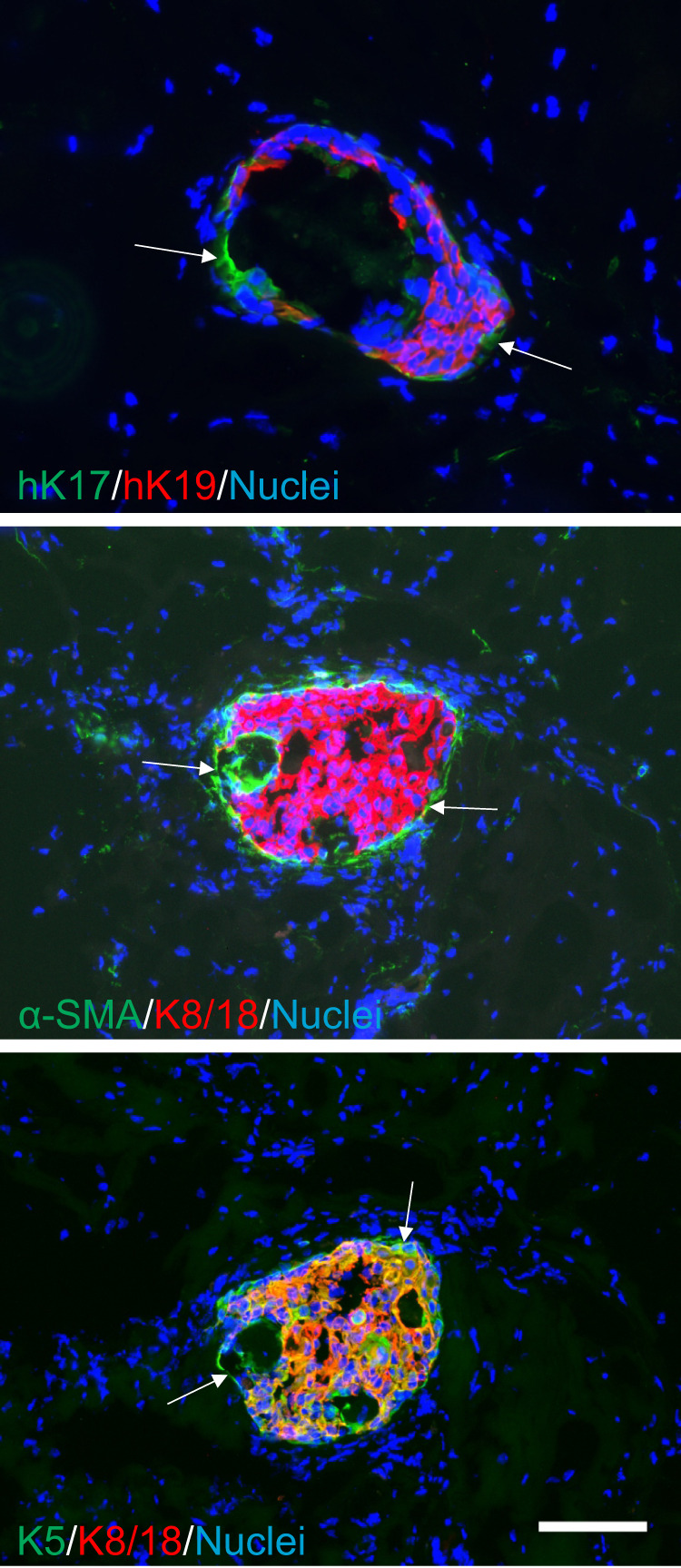
Fig. 6. Xenografted PIK3CA transformed CD200low MEP progenitors give rise to luminal hyperplastic lesions.

Fluorescence micrographs of structures formed by CD200low-hTERT-shp53-PIK3CAH1047R in vivo in NOG mice stained for human K17, (hK17), K5, α-SMA (green), human K19 (hK19), K8/18 (red), and nuclei (blue) show histology reminiscent of biphasic lesions with an outer layer of K17+, K5+, and α-SMA+ MEP cells (arrows), surrounding a core of hK19+/K5+/K8/18+ hyperplastic cells (scale bar, 100 μm).
