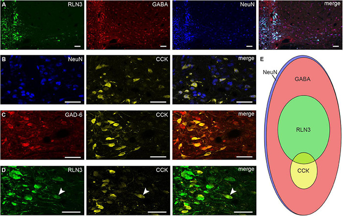
FIGURE 3.

RLN3, CCK, and GABA neurons in the nucleus incertus. Representative images of single coronal sections at the level of the NI illustrating: (A) RLN3 + (green), GABA + (red), NeuN + (blue) neurons and merged image; (B) NeuN + (blue), CCK + (yellow) cells and merged image; (C) GAD-6 + (red), CCK + (yellow) neurons and merged image; (D) RLN3 + (green) and CCK + (yellow) neurons, and sparse peptide colocalization (white arrowhead). Scale bars: 50 μm. (E) Schematic of the proportions of and relationship between all, RLN3 +, CCK +, and GABAergic NI neurons (area of each ellipse matches the percentage of each specific cell type). CCK, cholecystokinin; GABA, γ-aminobutyric acid; GAD-6, glutamic acid decarboxylase 65/67; NeuN, neuronal nuclear protein; RLN3, relaxin-3.
