FIGURE 5.
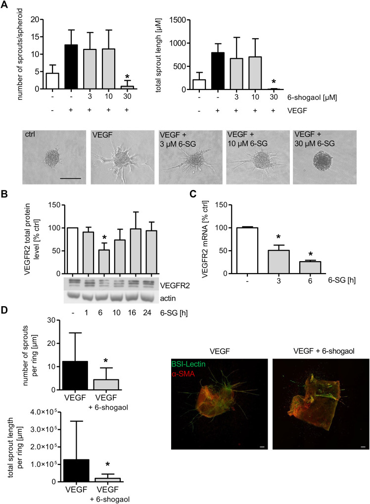
Formation of endothelial sprouts from HUVEC spheroids or mouse aortic rings is significantly attenuated by 6-shogaol. (A) HUVEC spheroids comprised 400 cells that were embedded into a rat tail collagen I gel. The spheroids were treated with indicated concentrations of 6-shogaol for 30 min before endothelial sprouting was induced by VEGF (10 ng/ml). 20 h later, the spheroids were fixed with 4% formaldehyde, and microscopic images were taken. Image quantification was performed using ImageJ. Scale bar represents 100 µm. One representative image for each condition is shown. (B,C) Confluent HUVECs were treated with 6-shogaol (30 µM) for indicated time points. (B) Total protein levels of VEGFR2 were determined by Western blot analysis. Actin served as the loading control. One representative blot is shown. (C) VEGFR2 mRNA data were obtained by qPCR. GAPDH served as the housekeeping gene. (D) Mouse aortic rings were embedded into a rat tail collagen I gel and stimulated with VEGF (30 ng/ml). After the formation of initial sprouts, the aortic rings were treated with 6-shogaol (30 µM) or vehicle (0.03% DMSO) for 3 days. Aortic rings were fixed and stained with BS-I lectin (green) and for α-smooth muscle actin (red). Laser confocal scanning microscopy and ImageJ allowed the quantification of sprout number and length. Scale bar represents 100 µm, and one representative image is shown. 6-SG: 6-shogaol. Data are expressed as mean ± SD; (A) n = 3; *p ≤ 0.05 vs. VEGF; (B) n = 3; (C) n = 3; *p ≤ 0.05 vs. ctrl; (D) n = 3; p ≤ 0.05 vs. VEGF.

