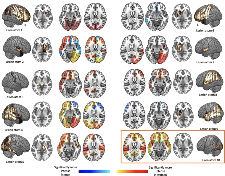
Figure 4.
Significantly altered male- and female-connectome-based lesion connectivity in 14 cortical networks (seven per hemisphere). Sex-specific lesion connectivity was computed for each of the 10 unique lesion patterns (c.f., Fig. 1) and subsequently statistically compared within each of the cortical networks. Networks with significantly different lesion connectivity are represented in colour (orange/red: significantly stronger in women; blue: significantly stronger in men). To allow for these statistical group comparisons in the first place, we inserted an additional simulation step: each lesion pattern was slightly varied 100 times by sampling a random number and collection of lesion pattern-affiliated brain regions. Exemplarily more in detail: Lesion pattern #1 primarily comprised the parcels pre- and post-central gyrus, superior, middle or inferior frontal gyrus, insular cortex, superior parietal and supramarginal cortex. We would here, for example, randomly choose pre- and post-central gyrus for a first Lesion pattern #1-like lesion, subsequently choose superior parietal, as well as supramarginal cortex for a second Lesion pattern #1-like lesion and so forth until we obtained 100 Lesion pattern #1-like lesions. Male- and female-specific lesion connectivity was then computed for each of these 100 simulated lesions per lesion pattern. Lesion patterns #2 and #10 comprised most connectivity differences (nine each). While Lesion pattern #2 was characterized by both higher connectivity in men and women, Lesion pattern #10 featured higher lesion connectivity exclusively in women.

