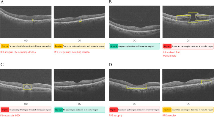
Figure 3.
Correct OCT-AI analysis reports of four participants. (A) Routine case in which RPE irregularity including drusen was found in both eyes. (B) Urgent case as macular hole and intraretinal fluid are detected in the left eye. (C) Another urgent case with fibrovascular PED detected in the right eye. (D) A case also predicted as urgent: RPE atrophy is found in both eyes and the right eye is considered an urgent level. Note that among eight OCT images per eye, one image containing typical pathologies or passing through the fovea is selected for display in the report.

