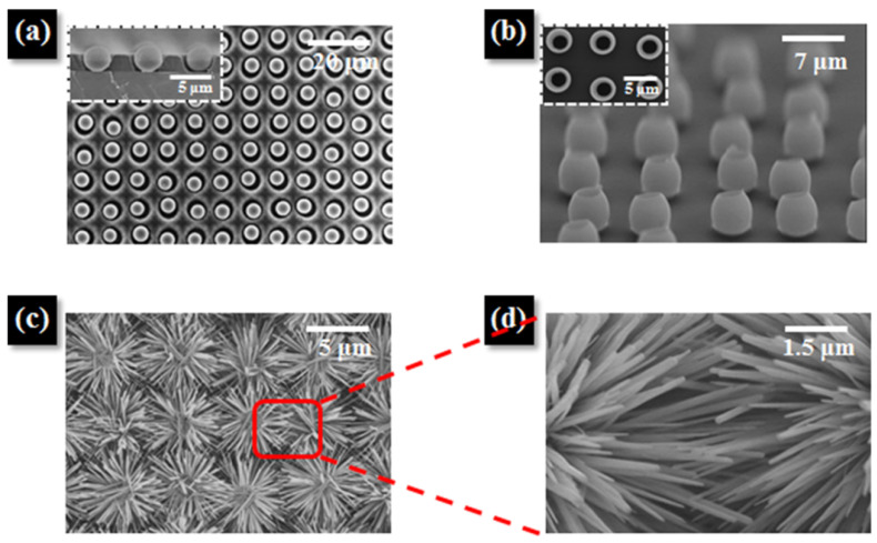
Figure 10.
(a) Top-view SEM image of a microparticle array in circular well patterns. Inset: Cross-sectional SEM image showing microparticles arrayed in height-optimized well patterns. (b) Tilted-view SEM image of aligned ZnO shells after transfer and calcination at 250 °C for 3 h in air on a hot plate. Inset: Top-view SEM image showing polystyrene-removed ZnO shells after calcination. (c) SEM image of the ZnO nanoflower network structure after growth of ZnO NRs in- and outside the shells, and (d) magnified SEM image of the junctions between the NRs. Reprinted with permission from Ref. [96]. Copyright 2017 American Chemical Society.

