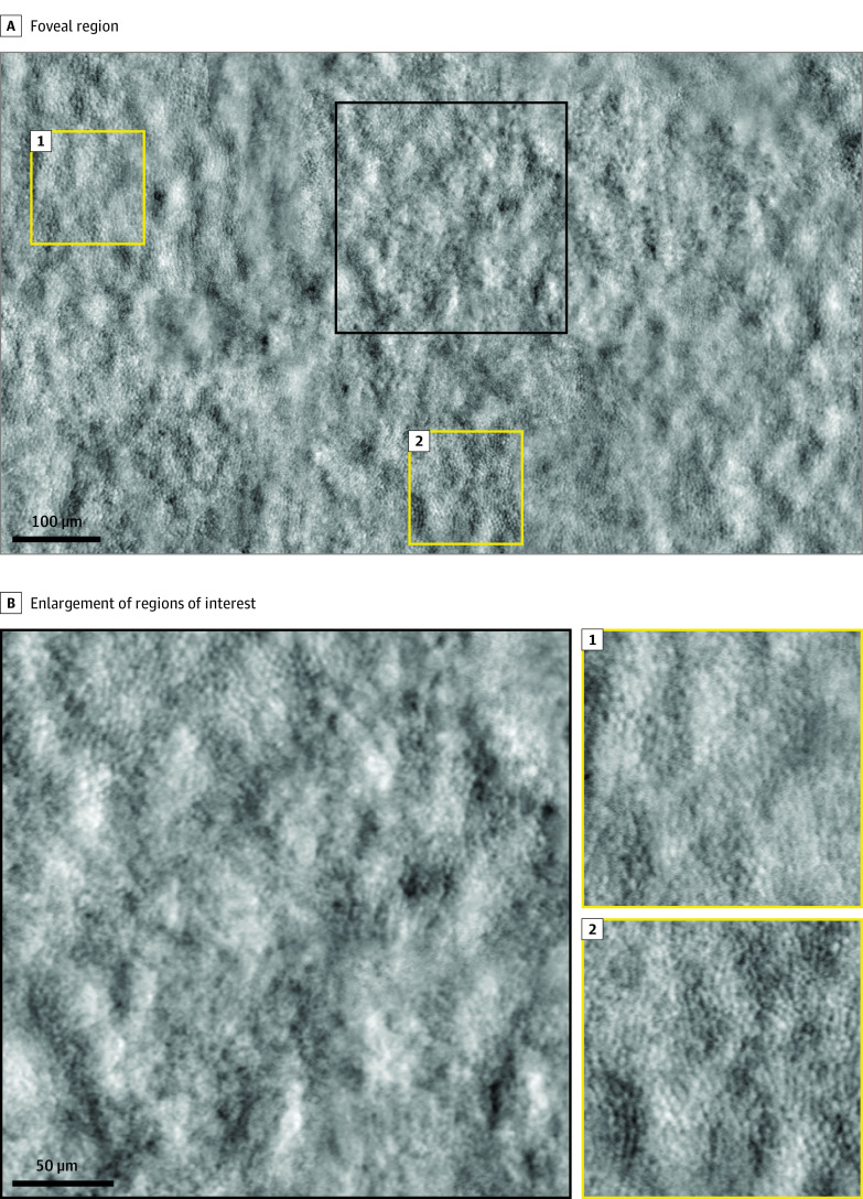
Figure 3. Adaptive Optics Nonconfocal Split Detection Montage in the Injected Eye of Patient 09 at 1 mo Postinjection.
The black box shows an area of loss of cone inner segments surrounded by intact cone inner segments at the fovea; yellow boxes show the intact cone inner segment mosaic in parafoveal regions.

