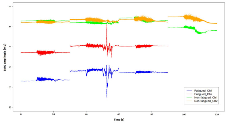
Figure 7.
Surface EMG signals of the left vastus lateralis (shown as Ch1) and the left rectus femoris (shown as Ch2) of a fatigued participant, compared with those of a participant who did not report fatigue. An epoch of 30 s EMG signals is extracted for each 10 s prompted contraction of the thigh, together with 10 s before and after the muscle contraction. The fourth muscle squeeze for the fatigued participant was not performed due to early termination of the head-up tilt test.

