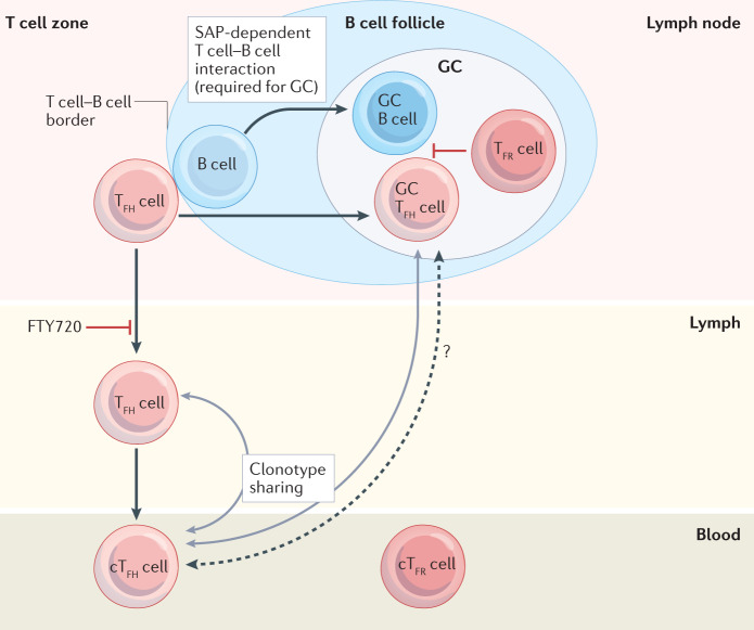
Fig. 1. Blood-borne cTFH cells are clonally related to their lymphoid counterparts.
Follicular helper T (TFH) cells classically reside in secondary lymphoid organs where they support germinal centre (GC) B cell responses; however, T cells with similar markers can be detected in the lymph and blood. These circulating TFH (cTFH) cells have a memory phenotype and do not depend on GCs as they can arise in the context of SLAM-associated protein (SAP) deficiency, which abrogates sustained T cell–B cell interactions22. However, they do require B cells, as cTFH cell frequencies are greatly reduced in individuals with B cell deficiency16. Blocking lymph node exit, by treatment with FTY720, dramatically decreases cTFH cells in mice and humans18,26,27. Clonotype sharing reveals a developmental relationship between TFH cells found in the GC and the blood24,25 and those found in the lymph and the blood26, suggesting that T cell–B cell interactions at the T cell–B cell border give rise to both GC TFH cells and cTFH cells. It is possible that some GC TFH cells enter the circulation from GCs but this is likely to be rare21. Follicular regulatory T (TFR) cells also have circulating counterparts (cTFR); however, these have an immature phenotype and differentiate from regulatory T cells without the requirement for contact with B cells164.

