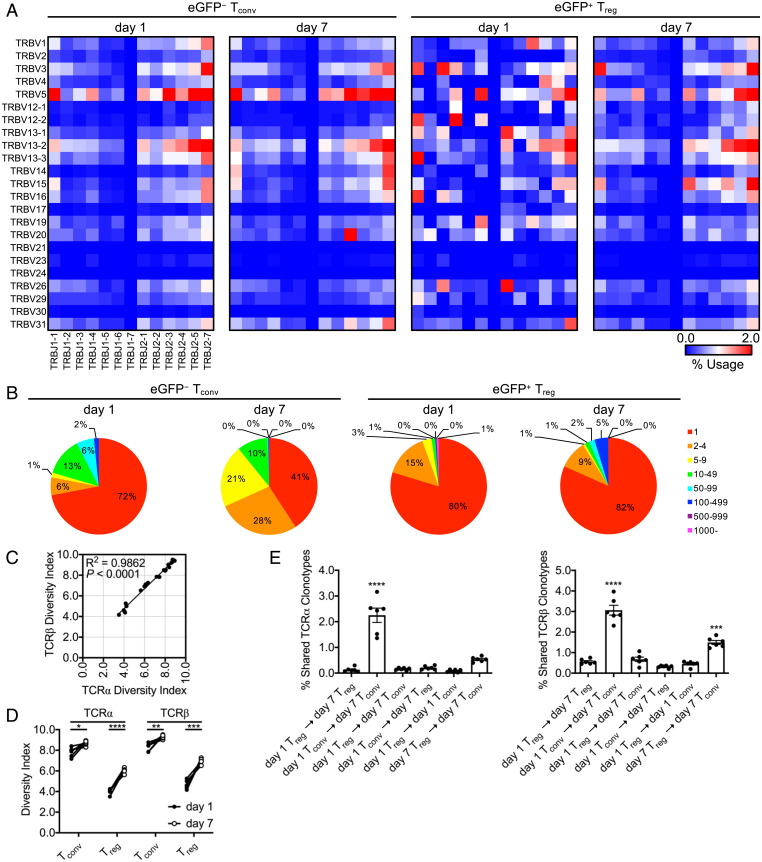
Fig. 2.
Splenic Tregs do not display clonal expansion during Listeria infection. Foxp3eGFP mice infected with L. monocytogenes ΔactA-Ova. Hemisplenectomy was performed at day 1 postinfection, and viable NK1.1−CD4+TCR-β+FoxP3-eGFP− (Tconv) and FoxP3-eGFP+ (Treg) cell populations were cell sorted from the dorsocranial lobe of the spleen. Corresponding cell populations were isolated at day 7 from the remaining ventral-caudal half of the spleen. Total RNA was isolated from all cells and subjected to TCR-Seq. (A) TCRβ Trb V (TRBV) and Trb J (TRBJ) gene segment family average usage among animals. (B) Representative number of Trb CDR3 clone reads. (C and D) Shannon–Weaver TCR diversity indices reported. (E) Intraanimal Venn analysis displaying shared Tra and Trb CDR3 clonotypes (n = 6 per group, 3 male and 3 female). Mean ± SEM. (C) R2/P (linear regression analysis); (D) *P < 0.05, **P < 0.01, ***P < 0.001, and ****P < 0.0001 (Student’s t test); and (E) ***P < 0.001 and ****P < 0.0001 (one-way ANOVA).

