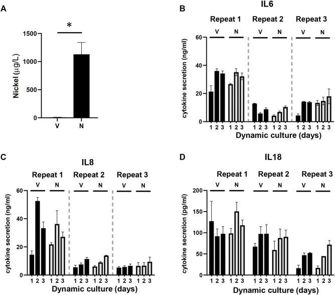
FIGURE 4.
Detection of nickel ions and interleukins. (A) Detection of the amount of nickel ions in culture supernatants derived from HUMIMIC Chip3plus, measured with graphite furnace atomic absorption spectrometry (GFAAS) in both vehicle (V)- and nickel (N)-exposed co-cultures at day 3. (B-D) Quantification of IL-6 (B), IL-8 (C), and IL-18 (D) in supernatants of multi-organ cultures upon exposure to the vehicle (V) or NiSO4 (N) over 3 days of culturing. The data represent mean ± SEM; n = 3. *, p < 0.05; paired t-test (A), 2-way ANOVA followed by Bonferroni’s multiple comparison test (B–D).

