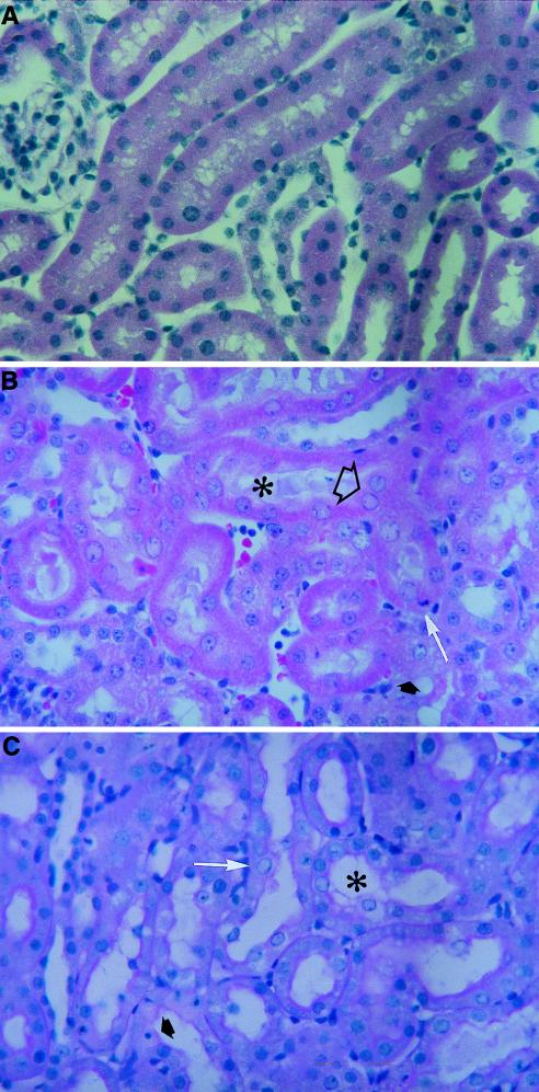
FIG. 6.
Histopathology analysis of kidneys from rats treated with cidofovir and compound 1. The animals were administered a solution of saline (A), a dose of 100 mg of cidofovir per kg (B), or a dose of 100 mg of compound 1 per kg (C) subcutaneously once daily for 5 consecutive days. (B) Cytoplasmic changes included effacing of the fine structural details (➧) and shedding into the lumen (✻). Nuclear changes consisted of karyomegaly and eosinophilic pseudoinclusions (➱) and apoptosis (➞). (C) Cytoplasmic changes included mild vacuolation and perinuclear hydropic degeneration (✻). Nuclear changes consisted of karyomegaly (➞) and apoptosis (➧).

