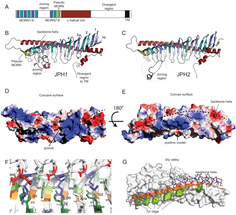
Fig. 1.
Crystal structure of JPH1 and JPH2 MORN-helical domain. (A) The primary sequence organization of JPH1-4 showing the 8 MORN repeats interrupted by a joining region, a pseudo-MORN repeat preceding a long α-helical–rich stretch, followed by a highly divergent region and ending in a short transmembrane helix. (B and C) The crystal structures of JPH1 and JPH2 (PDBs code 7RW4 and 7RXE) showing the MORN repeats, shortened joining region, and a long backbone helix. The conformation of the shortened joining region differs between the two isoforms. (D and E) The surface electrostatic potentials generated for JPH2 minus the shortened joining region, calculated in PyMOL, reveals a highly charged groove formed by the MORN repeats and an electropositive cluster at the center of the backbone helix. (F) A representation of the consensus MORN repeat-residues, including the Y1 and W5 consensus residues forming hydrophobic walls, accommodating R10, Q10, or K10 side chains. (G) The G3/G14 MORN residues forms a glycine valley, lined on one end by a tyrosine ridge formed by Y13 in some MORN repeats. The backbone helix fits in this valley using short alanine side chains.

