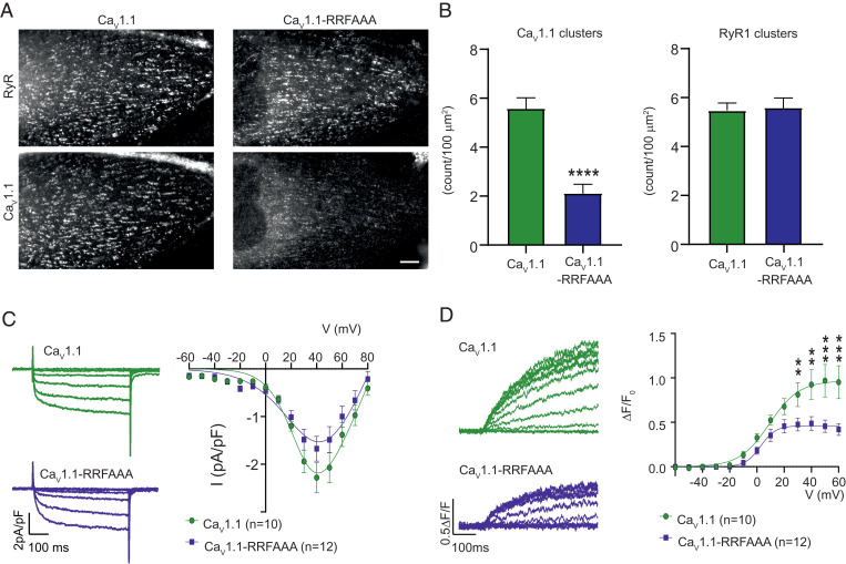
Fig. 3.
The RRFAAA mutation results in reduced junctional localization and reduced EC coupling. (A) Double immunofluorescence labeling of CaV1.1 and RyR1 shows that the RRFAAA mutation results in reduced incorporation of CaV1.1 in the T-tubule/SR junctions of dysgenic myotubes. (Scale bar, 10 µm.) (B) CaV1.1 cluster count reveals a significant (n = 30, Student’s t-test ****P < 0.0001) decrease of CaV1.1-RRFAAA incorporation in clusters compared to CaV1.1, while the clustering of RyR1 is unchanged (n = 30, Student’s t-test P = 0.83). (C) Representative calcium currents (Vtest at −40, −20, 0, +20, +40 mV, Left) and average IV relationships (Right) in dysgenic myotubes show a modest but statistically not significant effect of the RRFAAA mutation on L-type currents [two-way repeated-measures ANOVA F(1, 14) = 1.706; P = 0.054]. (D) Representative whole-cell voltage-clamp measurements of calcium transients with Fluo-4 (Left) and average peak change in fluorescence normalized by baseline (ΔF/F) as a function of test potential (Right) show a significant reduction of EC coupling calcium release. [Two-way repeated measures ANOVA F(1, 12) = 3.831; P < 0.001. The asterisks in the figure refer to the post hoc analysis: at +30 mV **P = 0.004, at +40 mV **P = 0.001, at +50 mV and +60 mV ***P < 0.001.] Error bars correspond to SEMs in B–D. Details of the fits are indicated in Materials and Methods.

